Pain stays focused
A specific area remains tender or painful and the underlying structure needs clarification.
 Ashleigh E. Korves, DPMFoot & Ankle CareESCall707-575-6033
Imaging & orthotics
An image can clarify structure, and support can change how pressure or motion is managed. Neither should be automatic. Both should connect to the diagnosis and what you need your feet to do.
Choosing useful tools
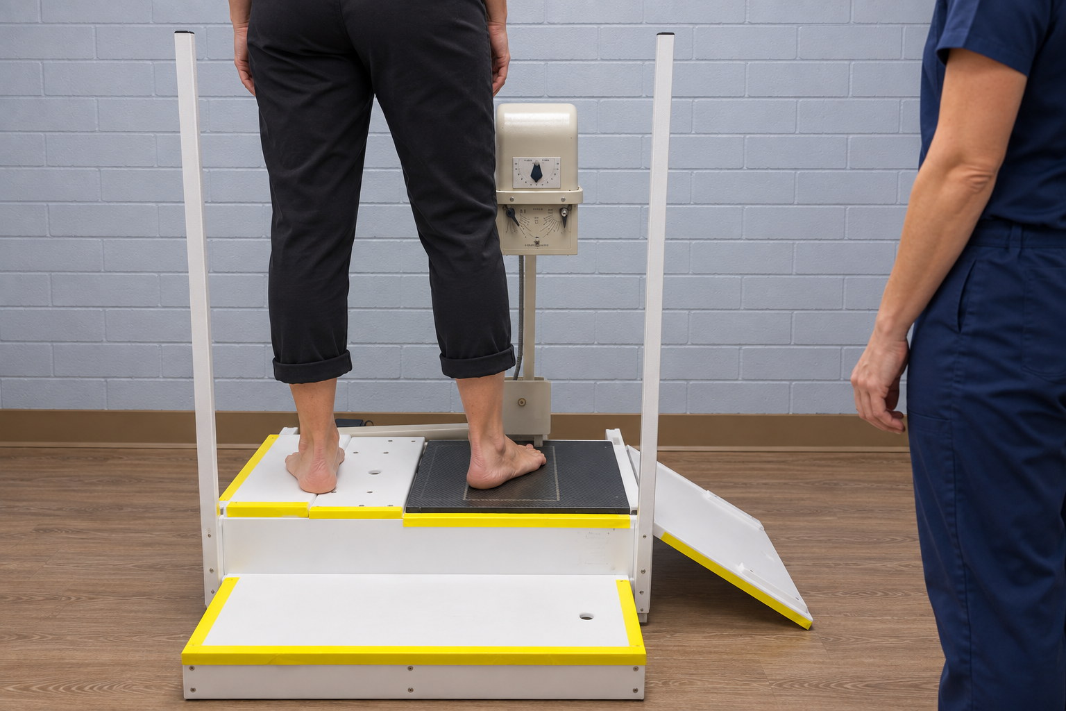
A clinical examination often provides the first information about tenderness, motion, strength, alignment, and function. Digital X-rays can add a view of bone and joint structure when that information may answer a specific question. Advanced imaging may be considered selectively rather than as a routine step for every symptom.
Support works the same way: footwear changes, over-the-counter inserts, padding, bracing, or custom orthotics should have a defined purpose. The most elaborate device is not automatically the right one. Comfort, activity, pressure, movement, and response over time help judge whether support is doing its job.
What you may notice
Persistent pain, alignment concerns, pressure points, or symptoms that may benefit from a clearer look at structure or support. The timing and location of symptoms are often as useful as the intensity.
A specific area remains tender or painful and the underlying structure needs clarification.
The way a foot or ankle stands or moves may be contributing to symptoms or shoe difficulty.
Callus, irritation, or another pressure point repeatedly develops in the same location.
Shoes, inserts, or an existing orthotic feel uncomfortable or have not changed symptoms as expected.
How Dr. Korves may help
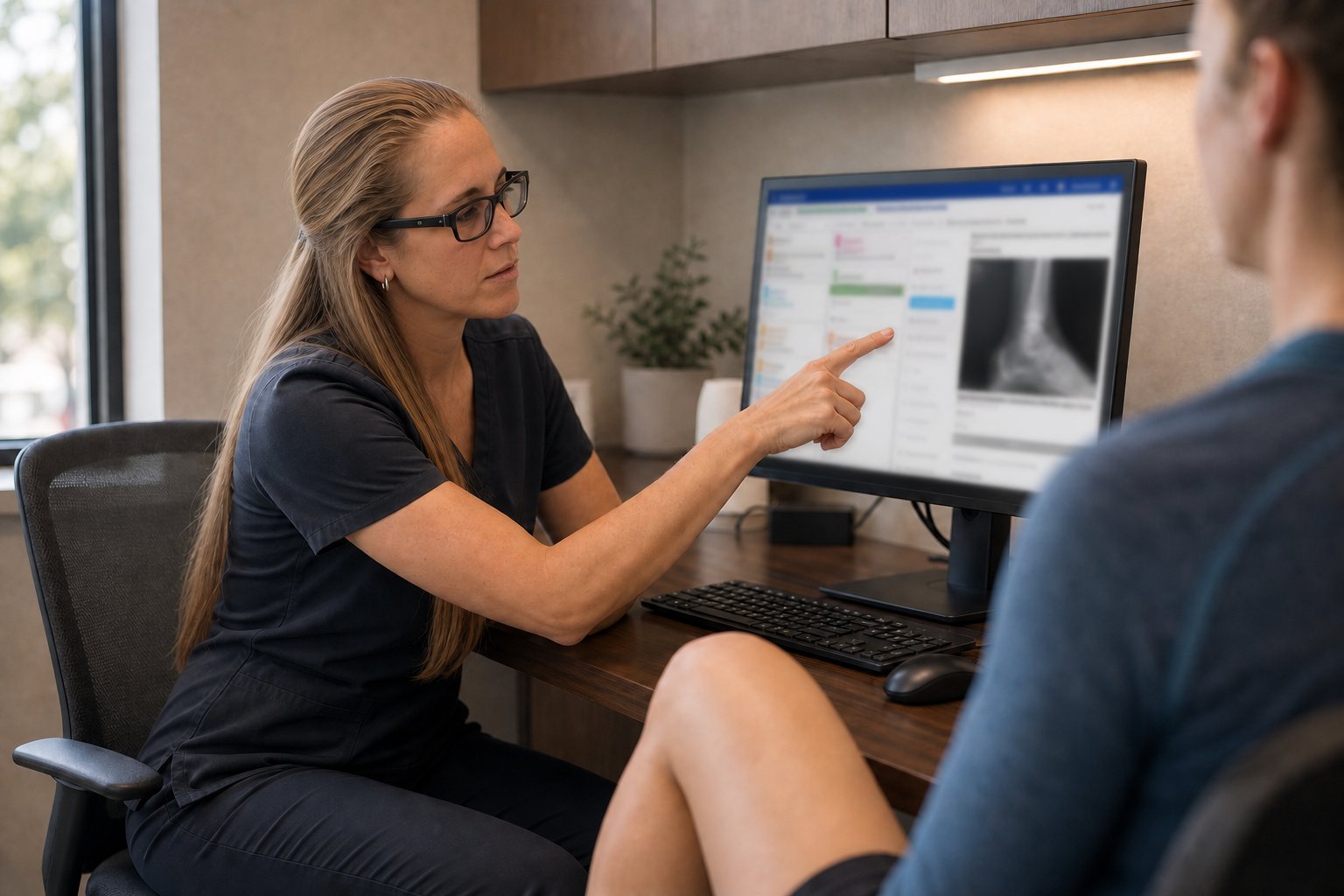
Dr. Korves begins with the symptom pattern and examination. If an image could clarify bone, joint, alignment, injury, or another structural question, she can explain why a digital X-ray or referral for different imaging may be useful.
When support is considered, the evaluation may include standing, walking, shoes, pressure, and the demands of work or activity. The recommendation can then distinguish a footwear change or readily available insert from a custom device or another approach.
Treatment possibilities
The goal may be to redistribute pressure, improve comfort, protect a painful area, or support a particular movement. The choice should be understandable and its effect should be reviewed.
Use an appropriate study when structural information is likely to refine the diagnosis or treatment decision.
Fit, shape, cushioning, stiffness, and activity-specific design may address some problems without a separate device.
Dr. Korves may recommend padding, inserts, bracing, or custom orthotic fabrication to address a specific need based on your diagnosis and activity.
Follow-up can assess fit, skin pressure, wear, symptom response, and whether an adjustment is needed.
Advanced options, when appropriate
Further imaging, bracing, rehabilitation, an office-based procedure, or a surgical consultation may be considered when symptoms and findings warrant it. Escalation should follow a clinical question rather than happen automatically because an X-ray or insert did not solve the problem.
Explore conservative care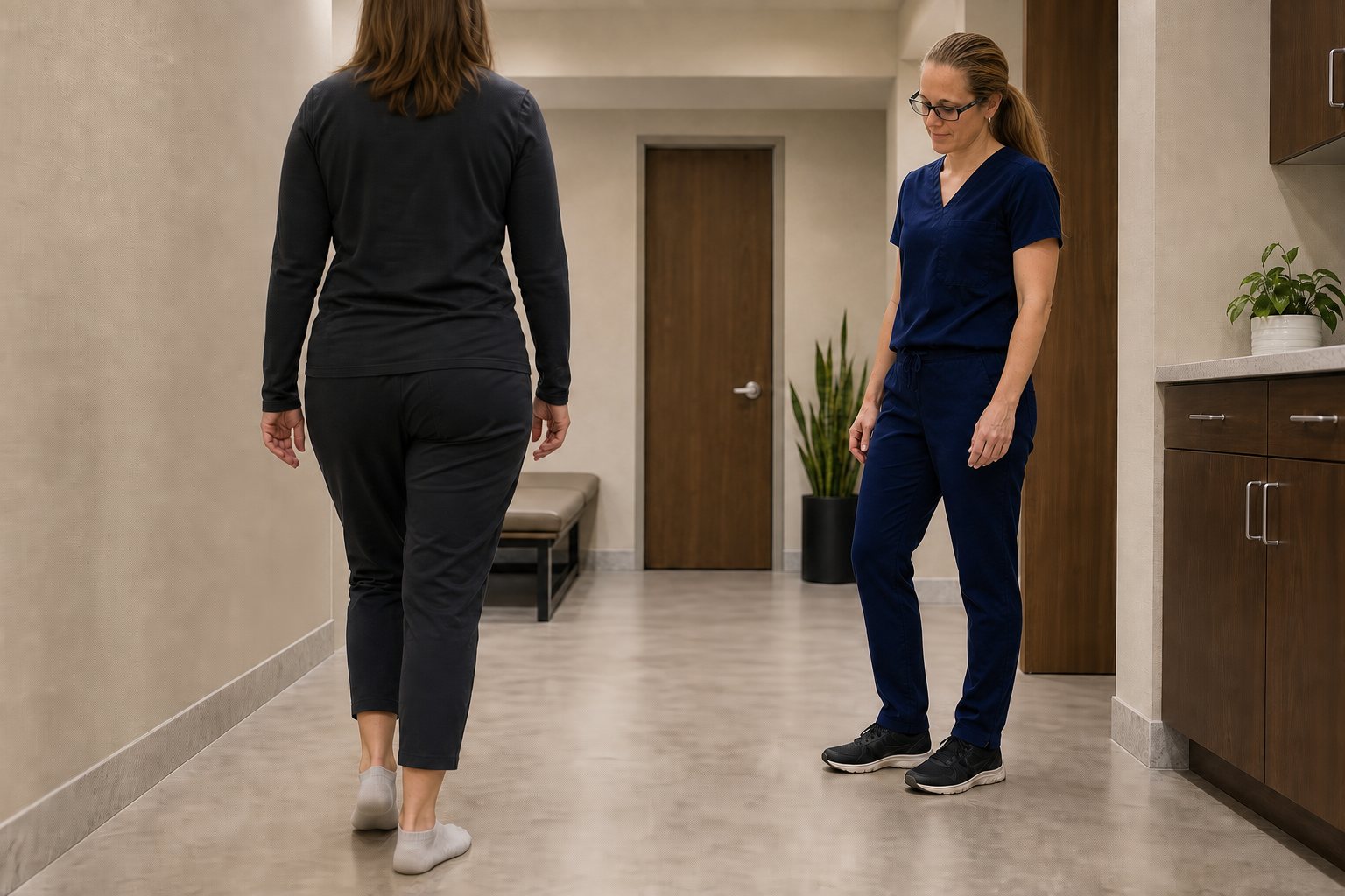
When to be seen
A significant injury, visible deformity, or inability to bear weight needs prompt evaluation rather than waiting for an orthotic visit.
Prompt care is also important for new loss of sensation or circulation, spreading infection signs, or an open area on a higher-risk foot.
For your appointment
What question would imaging answer?
Would an insert or custom orthotic change the mechanics that matter?
Which shoes should I bring?
How will we judge whether support is helping?
Choose with purpose
Call the office to discuss persistent symptoms, prior imaging, or support that is not working. An evaluation can help connect the right information to the right next step.